TSFM-HMGA2 Fusion FISH Probe
The TSFM-HMGA2 Fusion FISH Probe is used to confirm a fusion of the TSFM and HMGA2 genes. The fusion of the TSFM and HMGA2 genes has been associated with Skin Cutaneous Melanoma. These probes are FISH confirmed on normal peripheral blood in both interphase nuclei and metaphase spreads before shipment. Typical turnaround time for this product is 7-14 days after purchase.
** This product is for in vitro and research use only. This product is not intended for diagnostic use. Please note that both genes fall on the same chromosome and inter-chromosomal detection may be difficult to detect depending on the genes proximity to one another. Please consult our support staff before ordering this product to ensure that the probe can be designed to meet your specific needs.
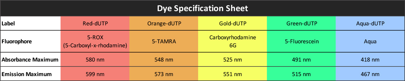
| SKU | Test Kits | Buffer | Dye Color | Order Now |
|---|---|---|---|---|
| TSFM-HMGA2-20-ORGR (Standard Design) | 20 (40 μL) | 200 μL |


|
|
| TSFM-HMGA2-20-RERE | 20 (40 μL) | 200 μL |

|
|
| TSFM-HMGA2-20-REOR | 20 (40 μL) | 200 μL |
|
|
| TSFM-HMGA2-20-REGO | 20 (40 μL) | 200 μL |

|
|
| TSFM-HMGA2-20-REGR | 20 (40 μL) | 200 μL |
|
|
| TSFM-HMGA2-20-REAQ | 20 (40 μL) | 200 μL |

|
|
| TSFM-HMGA2-20-ORRE | 20 (40 μL) | 200 μL |
|
|
| TSFM-HMGA2-20-OROR | 20 (40 μL) | 200 μL |
|
|
| TSFM-HMGA2-20-ORGO | 20 (40 μL) | 200 μL |
|
|
| TSFM-HMGA2-20-ORAQ | 20 (40 μL) | 200 μL |
|
|
| TSFM-HMGA2-20-GORE | 20 (40 μL) | 200 μL |
|
|
| TSFM-HMGA2-20-GOOR | 20 (40 μL) | 200 μL |
|
|
| TSFM-HMGA2-20-GOGO | 20 (40 μL) | 200 μL |
|
|
| TSFM-HMGA2-20-GOGR | 20 (40 μL) | 200 μL |
|
|
| TSFM-HMGA2-20-GOAQ | 20 (40 μL) | 200 μL |
|
|
| TSFM-HMGA2-20-GRRE | 20 (40 μL) | 200 μL |
|
|
| TSFM-HMGA2-20-GROR | 20 (40 μL) | 200 μL |
|
|
| TSFM-HMGA2-20-GRGO | 20 (40 μL) | 200 μL |
|
|
| TSFM-HMGA2-20-GRGR | 20 (40 μL) | 200 μL |
|
|
| TSFM-HMGA2-20-GRAQ | 20 (40 μL) | 200 μL |
|
|
| TSFM-HMGA2-20-AQRE | 20 (40 μL) | 200 μL |
|
|
| TSFM-HMGA2-20-AQOR | 20 (40 μL) | 200 μL |
|
|
| TSFM-HMGA2-20-AQGO | 20 (40 μL) | 200 μL |
|
|
| TSFM-HMGA2-20-AQGR | 20 (40 μL) | 200 μL |
|
|
| TSFM-HMGA2-20-AQAQ | 20 (40 μL) | 200 μL |
|
HMGA2 Gene Summary
This gene encodes a protein that belongs to the non-histone chromosomal high mobility group (HMG) protein family. HMG proteins function as architectural factors and are essential components of the enhancesome. This protein contains structural DNA-binding domains and may act as a transcriptional regulating factor. Identification of the deletion, amplification, and rearrangement of this gene that are associated with myxoid liposarcoma suggests a role in adipogenesis and mesenchymal differentiation. A gene knock out study of the mouse counterpart demonstrated that this gene is involved in diet-induced obesity. Alternate transcriptional splice variants, encoding different isoforms, have been characterized. [provided by RefSeq, Jul 2008]
Gene Name: High Mobility Group AT-hook 2
Chromosome: CHR12: 66218239 -66360071
Locus: 12q14.3
TSFM Gene Summary
This gene encodes a mitochondrial translation elongation factor. The encoded protein is an enzyme that catalyzes the exchange of guanine nucleotides on the translation elongation factor Tu during the elongation step of mitchondrial protein translation. Mutations in this gene are associated with combined oxidative phosphorylation deficiency-3 syndrome. Alternate splicing results in multiple transcript variants.[provided by RefSeq, Mar 2010]
Gene Name: Ts Translation Elongation Factor, Mitochondrial
Chromosome: CHR12: 58176527 -58196639
Locus: 12q14.1
Gene Diseases
The TSFM HMGA2 Fusion has been associated with the following diseases:
| Disease Name |
|---|
| Skin Cutaneous Melanoma |
FISH Probe Protocols
| Protocol, Procedure, or Form Name | Last Modified | Download |
|---|