EBER CISH Kit
$1,600.00
Not available in the USA.
Description
The EBER CISH Kit from Empire Genomics is designed for the detection of Epstein–Barr virus (EBV)–encoded RNA (EBER) in formalin-fixed, paraffin-embedded (FFPE) tissue sections using chromogenic in situ hybridization (CISH). This assay is optimized for automated workflows on the NeoPath Pro Automated Slide Stainer, enabling consistent processing and visualization of EBV-infected cells directly within tissue morphology using standard brightfield microscopy.
The kit contains a probe targeting EBER1 and EBER2 RNA, two highly abundant non-coding RNAs expressed during latent infection with Epstein–Barr virus. Because these transcripts are present at very high levels in infected cells, they provide reliable molecular targets for in situ EBV detection in tissue samples.
After hybridization of the digoxigenin-labeled probe to complementary EBER RNA sequences, a peroxidase-based chromogenic detection system with DAB produces a visible reaction product at the hybridization site. This signal marks the location of EBV RNA within the tissue section while maintaining cellular architecture. Slides can then be counterstained and examined using a standard light microscope.
Detection of EBER RNA in Tissue
Epstein–Barr virus (EBV) is a widely prevalent human herpesvirus that establishes lifelong latency following infection. During latency, EBV persists primarily in B lymphocytes and expresses a limited set of viral genes, including EBER1 and EBER2 RNAs. These transcripts are the most abundant viral RNAs found in latently infected cells and are highly conserved across EBV strains.
Because of their abundance and stability, EBER transcripts are commonly used targets for detecting EBV within tissue sections using chromogenic in situ hybridization (CISH). This approach enables researchers and pathology laboratories to localize EBV-infected cells while preserving the histological context of the specimen.
Automated CISH Workflow
The EBER CISH Kit for NeoPath Pro supports automated chromogenic in situ hybridization workflows. Within the automated protocol, the labeled probe hybridizes to complementary EBER RNA sequences present in infected cells. An enzyme-linked detection system then generates a chromogenic signal that can be visualized using brightfield microscopy.
Automation with the NeoPath Pro helps laboratories achieve standardized reagent delivery, consistent hybridization conditions, and reproducible staining results across tissue samples.
Kit Components
| Component | Volume |
|---|---|
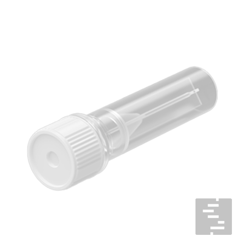
| EBER Probe | 1 × 1.41 mL |
| Digoxigenin CISH Detection Reagent | 1 × 7 mL |
| Proteinase K | 1 × 14 mL |
Kit Size: 20 Tests
Key Features
-
Designed for EBER RNA detection in FFPE tissue sections
-
Optimized for automated workflows on the NeoPath Pro
-
Uses chromogenic in situ hybridization (CISH) for brightfield visualization
-
Targets EBER1 and EBER2 transcripts expressed during latent EBV infection
-
Preserves tissue morphology while detecting viral RNA within cells
Certificate of Analysis (CoA)
Please check back later for your digital copy, contact us today for your CoA.
Documentation

Customer Publications
Reviews
Related products
Break Apart
Concentrate

Concentrate
Concentrate
Concentrate
Ancillary
Concentrate
CE-IVD
Reviews
There are no reviews yet.