SEARCH OUR PRODUCT CATALOG

HMGA2 Break Apart FISH Probe

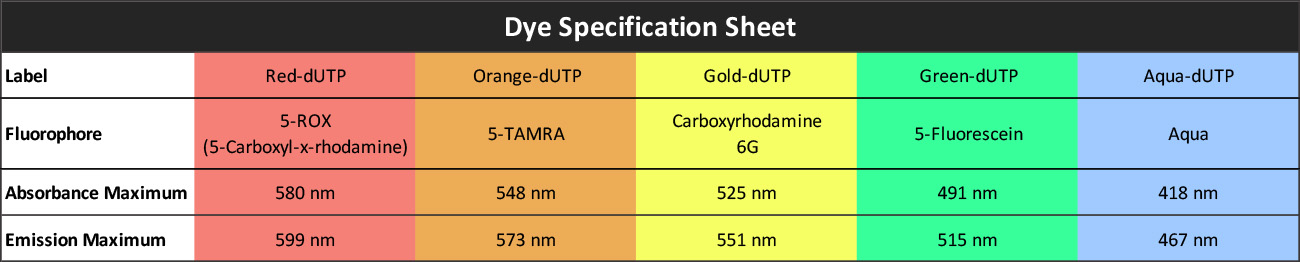
Our HMGA2 Break Apart Probe is designed to detect HMGA2 translocations. The probe comes labeled in orange and green, but can be customized to meet your needs.

** This product is for in vitro and research use only. This product is not intended for diagnostic use.

Turnaround Time: 7-10 Business Days    Shipping Time: 1-2 Day Expedited Shipping

SKU Test Kits Buffer Dye Color Order Now
HMGA2BA-20-ORGR  (Standard Design) 20 (40 μL) 200 μL
HMGA2BA-20-GRRE 20 (40 μL) 200 μL
HMGA2BA-20-GROR 20 (40 μL) 200 μL
HMGA2BA-20-REGR 20 (40 μL) 200 μL

Gene Summary

This gene encodes a protein that belongs to the non-histone chromosomal high mobility group (HMG) protein family. HMG proteins function as architectural factors and are essential components of the enhancesome. This protein contains structural DNA-binding domains and may act as a transcriptional regulating factor. Identification of the deletion, amplification, and rearrangement of this gene that are associated with myxoid liposarcoma suggests a role in adipogenesis and mesenchymal differentiation. A gene knock out study of the mouse counterpart demonstrated that this gene is involved in diet-induced obesity. Alternate transcriptional splice variants, encoding different isoforms, have been characterized. [provided by RefSeq, Jul 2008]

Gene Details

Gene Symbol: HMGA2

Gene Name: High Mobility Group AT-hook 2

Chromosome: CHR12: 66218239-66360071

Locus: 12q14.3

FISH Probe Protocols

Protocol, Procedure, or Form Name Last Modified Download

Epithelial-Myoepithelial Carcinoma Frequent Morphologic and Molecular Evidence of Preexisting Pleomorphic Adenoma, Common HRAS Mutations in PLAG1-intact and HMGA2-intact Cases, and Occasional TP53, FBXW7, and SMARCB1 Alterations in High-grade Cases

There is though to be a relationship between genetic aberrations, preexisting pleomorphic adenoma (PA), and the structural abnormality of epithelial-myoepithelial carcinomas (EMCAs). EMCAs were analyzed on a molecular level for PA by using FISH. FISH analysis was used with our break apart probes to detect PLAG1 and HMGA2 rearrangements. It was found that a relationship was present, as 80% of EMCA arise from PA, and EMCA genetics can vary with the status of PLAG1 and HMGA2.

Recurrent rearrangements of the PLAG1 and HMGA2 genes in lacrimal gland pleomorphic adenoma and carcinoma ex pleomorphic adenoma

Lacrimal gland tumors are histologically similar to salivary gland tumors. In the salivary glands, pleomorphic adenoma (PA) and carcinoma ex pleomorphic adenoma (ca_ex_PA) are both characterized by PLAG1 and HMGA2 gene rearrangements. However, it is not known if these rearrangements are present in lacrimal gland PA and ca_ex_PA. FISH analysis was done with our PLAG1, HMGA2, and NFIB break apart probes. It was found that rearrangements were frequent in the tested lacrimal glands and that testing for the rearrangement can help in distinguishing lacrimal gland PA and ca_ex_PA from de novo carcinomas.

Hydropic leiomyoma: a distinct variant of leiomyoma closely related to HMGA2 overexpression

Hydropic leiomyoma (HLM) is an understudied subtype of uterine leiomyoma (ULM). Little is known about HLM's etiology, genetics, and clinical profile. To account for this lack of data, the team analyzed 24 HLM cases for histology, immunohistochemistry and molecular alterations. Empire Genomics’ HMGA2 break apart probe was used to detect rearrangements of HMGA2, an abnormality previously reported in ULM. The gene was found rearranged in 32% of cases.

Metastasizing Pleomorphic Adenoma: Recurrent PLAG1/HMGA2 Rearrangements and Identification of a Novel HMGA2-TMTC2 Fusion

Pleomorphic adenoma (PA), although considered benign, can undergo malignant transformation, and metastatic cases are well documented. There is a lack of research on genetic characterization of metastasizing versus benign PA. In this study, four cases of metastasizing PAs were cytogenetically profiled using both RNA sequencing and FISH. PLAG1 and HMGA2 break-apart probes from Empire Genomics were used to confirm rearrangements of the genes. PLAG1 rearrangements were detected in all four cases. Results demonstrated that MPA harbors the same disease-defining molecular hallmarks as their benign counterparts.

Giant Cell Carcinosarcoma of the Parotid Gland With a PLAG1 Translocation in Association With a Pleomorphic Adenoma With HMGA2 Translocation: A Case Report and Fluorescence In Situ Hybridization Study

This case study looked at a parotid gland carcinosarcoma in a 77 year old patient. Our PLAG1 and HMGA2 break apart probes were used to detect rearrangements of the genes in both the carcinomatous and sarcomatous components of his tumor, to determine whether they harbored different genetic lesions. Both components were positive for PLAG1 and negative for HMGA2 translocations.